
Occlusal Change Through Orthodontics in TMD Patients
Originally posted on Dentistry Today.
Although some claim that occlusion has little effect on a healthy TMJ and is not generally a causal factor in TMD,1,2 many have made emphatic claims to the contrary.3 Many orthodontic practices are positioned in the marketplace as providing proven treatment for TMD, yet some patients seem to experience TMD as a consequence of orthodontic treatment.
While existing literature reports that orthodontics can both help4 and worsen TMD, this paper describes 2 cases where TMD relief was successfully achieved through orthodontic therapy. More specifically, these cases lend credence to the theory that increasing the vertical dimension5 and removing retrusive forces on the mandible may help recapture the disc that can be displaced by over-closure of the mandible.
CASE 1
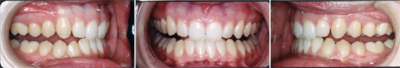 |
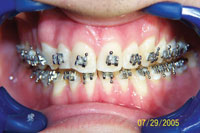
| Figure 1. Photos of patient when she presented for fixed orthodontic treatment. She had been wearing a removable splint and was asymptomatic but splint dependent. |
Case 1 describes treatment that utilized a hard acrylic, flat-planed splint to alleviate TMD symptoms of pain, popping, and clicking by advancing the mandible and increasing vertical dimension. The patient was “splint dependent” but symptom-free at the stage she was transferred for orthodontic treatment (Figure 1). The pain returned whenever she was not wearing her splint for consecutive days because she returned to an “over-closed” position. Once orthodontic treatment commenced, the splint was reduced incrementally, allowing teeth to supra-erupt. This was done sequentially until the natural occlusion mimicked the patientís occlusion with the splint. It was reduced from the posterior forward, allowing the second molars to supra-erupt in a controlled fashion. It was also sequentially reduced in thickness. Mobility from the orthodontics facilitated this occlusal setting. Three distinct aspects of the patientís occlusion were changed, which helped provide TMD relief:
(1) The maxillary incisors were flared labially with treatment. Lingually inclined lower incisors translate occlusal force into a retrusive direction as the patient closes, especially during protrusion. This was eliminated as labially inclined upper and lower incisors deliver chewing force in a more vertical direction into the alveolar bone, decreasing the tendency of the mandible to be pushed backward and minimizing disc trauma.
(2) Similarly, the incisors had greater vertical overlap initially. This compounded the problem caused by the retroclined position, as the entire facial surface of the lower incisors was acting as a receiving surface for ìpoundingî by the maxillary incisors. The posterior dentition better tolerates this vertical chewing force.
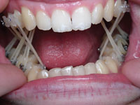
Figure 2. Cross-arch vertical elastics used to bring posterior extrusion without tipping. The splint was reduced incrementally. (3) The molar extrusion and improved interdigitation, in conjunction with occlusal adjustment, provided a more stable posterior occlusion. This offers better protection against retrusive slides in centric and during mastication, which can further exacerbate TMD. Molar extrusion achieved using cross-arch elastics (Figure 2) from the buccal of the upper teeth to the lingual of the lowers as well as lingual of the upper teeth to the buccal of the lowers served to extrude the posteriors with greater control and no buccal-lingual tipping.
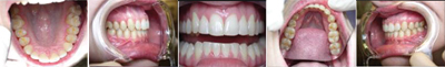 |
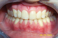
| Figure 3. Occlusion after removal of braces. |
Although the causal factors of TMD are often a mystery, this case demonstrates that eliminating obvious and severe occlusal abnormalities through splint therapy and gradually through or-thodontics may provide TMD relief and minimize occlusal wear as the traumatic occlusion is eliminated (Figure 3). Two years after treatment, the patient was orthodontically stable and symptom-free.
CASE 2
 |
 |
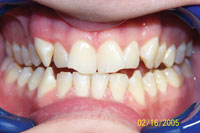
| Figure 4. Patient’s occlusion before treatment. | Figure 5. Progress at 5 months. |
 |
 |
| Figure 6. “After” photo with upper and lower teeth splinted and incisals restored. | Figure 7. Eleven-month recall. |
The second case shows a patient who had bilateral TMJ clicking and tinnitus. He had second molar occlusion only, a constricted maxillary arch, occlusal trauma, and wear (Figure 4).
The patient wore posterior cross-arch elastics from the lingual of the maxillary posteriors to the buccal of the mandibular posteriors to achieve proper intercuspation and bilateral, evenly distributed tooth contacts, as a posterior cross-bite has been associated with TMD.6 The upper posteriors were stabilized with a Hawley retainer. The upper and lower anteriors were stabilized with lingual Ribbond splints (Ribbond) canine to canine.
This effectively stabilized rotated teeth (in conjunction with a fiberotomy) and provided proper resistance form to the restored incisal composites, necessary because of the previous occlusal trauma (Figures 5 and 6). The incisal edges became much more durable once connected to the splint because of increased thickness. The TMJ, occlusion, and restored incisal surfaces were all stable at recall (Figure 7).
CONCLUSION
References
- Gesch D, Bernhardt O, Kirbschus A. Association of malocclusion and functional occlusion with temporomandibular disorders (TMD) in adults: a systematic review of population-based studies. Quintessence Int. 2004;35:211-221.
- Gesch D, Bernhardt O, Mack F, et al. Association of malocclusion and functional occlusion with subjective symptoms of TMD in adults: results of the Study of Health in Pomerania (SHIP). Angle Orthod. 2005;75:183-190.
- Reinhardt R, Tremel T, Wehrbein H, et al. The unilateral chewing phenomenon, occlusion, and TMD. Cranio. 2006;24:166-170.
- Henrikson T, Nilner M, Kurol J. Signs of temporomandibular disorders in girls receiving orthodontic treatment. A prospective and longitudinal comparison with untreated Class II malocclusions and normal occlusion subjects. Eur J Orthod. 2000;22:271-281.
- Hisano M, Ohtsubo K, Chung CJ, et al. Vertical control by combining a monoblock appliance in adult class III overclosure treatment. Angle Orthod. 2006;76:226-235.
- Thilander B, Rubio G, Pena L, et al. Prevalence of temporomandibular dysfunction and its association with malocclusion in children and adolescents: an epidemiologic study related to specified stages of dental development. Angle Orthod. 2002;72:146-154.
- Clark GT. Etiologic theory and the prevention of temporomandibular disorders. Adv Dent Res. 1991;5:60-66.