Straight Teeth in Six Months
Call Now for a Free Consultation: (617) 277-5200

Call Now for a Free Consultation: (617) 277-5200
Brookline rapid braces

All Posts in Category: Expert Boston Dental Care

Six-Month Adult Aesthetic Orthodontic Treatment

Six-Month Adult Aesthetic Orthodontic Treatment

Straight teeth in just six months.

Posted on Glidewell Laboratories.

While cosmetic dentistry has commanded more attention with recent breakthroughs — such as all-ceramic crowns, veneers, composite materials and intraoral cameras — the demand for adult cosmetic orthodontic treatment has also increased. It has been estimated that in 1970, only 5 percent of adults aged 18 or older sought consultations for comprehensive orthodontic treatment.1 In 1990, four times that number sought consultations for orthodontics.

Currently, adults present with chief complaints about the crowding of their teeth more frequently than anything else.2 Many adult patients want to straighten their teeth, but they are unwilling to wear braces for two or more years. Patients presenting with a physiologic occlusion and a desire for aesthetic improvement can benefit from orthodontic correction that requires only a short treatment time of six months or less. Adults who have their teeth straightened experience a better body self-image and higher self-esteem.3,4 The general public is focused on a noncrowded, aesthetic tooth arrangement more so than orthodontists, who are also concerned with occlusal and skeletal relations.5 A short, six-month treatment can very well enhance periodontal and occlusal aspects of the patient’s dentition. Treatment, therefore, serves as an adjunct to final periodontic and restorative treatment, even though the main focus remains cosmetic.

Simultaneously treatment planning the orthodontics with the cosmetics, crown & bridge, and periodontics in the same office facilitates a well-orchestrated cosmetic result, which can be more difficult to achieve through cross communicating between specialists. In this context, limited cosmetic orthodontic treatment is best done on patients who otherwise may not opt for comprehensive orthodontic treatment.

Method

The first aspect of case selection involves a discussion of the patient’s chief complaint. Patients are given a list of orthodontic and cosmetic problems, and asked to indicate their objective(s) for seeking treatment. In almost 90 percent of adult cases, relieving anterior crowding is the primary concern. This figure is based on 20 to 25 new orthodontic consults per month for six months in my general practice.

When the patient is committed to treatment, a database of information should be obtained: panoramic and full-mouth radiographs, intraoral and extraoral photographs, and models. A problem list is then reviewed with the patient, followed by a comprehensive treatment plan. The orthodontic aspect should be cosmetically oriented, specifically excluding skeletal problems. Because the profile and posterior occlusion are not to be changed significantly, a lateral cephalometric X-ray is not necessary.6

The treatment sequence includes the following:

  • Data collection and records;
  • Prophylaxis, fluoride application, oral hygiene instruction, and endodontic and periodontic disease resolution;
  • Extraction of third molars and a lower incisor when necessary (other teeth may rarely need to be extracted);
  • Cosmetic orthodontics; and
  • Bleaching, crowns and cosmetic bonding when indicated

If the patient prefers not to wear Hawley retainers, teeth can be retained by splinting once settling has occurred.

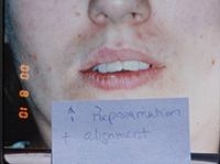
The Case for Enamel Reproximation

Because the postextraction health of the temporomandibular joint has been questioned, bicuspid extraction is now done with less frequency than in the past. It provides a result that is not always aesthetic or stable, has been slowly decreasing in popularity (almost 8 percent between 1988 and 1993), and remains controversial, varying widely among practitioners.7-10 Almost one and a half years is required to close the extraction spaces, and nonextraction patients have fuller lip support following treatment.11 Expansion is also a questionable method of treatment because long-term stability is doubtful.12

However, enamel reproximation allows for minimal localized tooth movements, fewer extractions, maintenance of lip support and shorter treatment time. Begg theorized that crowding of most dentitions is actually the result of decreased proximal wear, which our evolutionary predecessors once experienced.13 Therefore, enamel reproximation would seem to be the most natural available remedy for relieving crowding.

Enamel reproximation (air-rotor stripping) can be done for up to a 10 mm arch-length discrepancy. Sheridan recommends limiting reproximation to 1 mm per contact or 0.5 mm per proximal surface.14,15 Frequently, more than this can be done without noticeable change in tooth morphology or sensitivity because it’s done throughout six months in conjunction with fluoride treatments. It has also been theorized that the resultant flat interproximal contacts may actually increase post-treatment stability.16 Anterior lower arch crowding greater than 4 mm should be treated with the extraction of a lower incisor, followed by reproximation to minimize the black triangular space at the gumline. In most cases, a space determination is performed by resetting the teeth on the models with wax to measure the space required. This also allows a preview of the aesthetic result for both patient and doctor.

Appliances

Brackets should be bonded to the first molars using a straight-wire technique and NiTi wires. Posterior brackets with a larger (0.022) bracket slot placed in an ideal, aligned position minimize posterior occlusal changes. Successive reproximation using double-sided fine diamond discs (Brasseler) is followed by the use of fluted carbide burs for finishing and rounding enamel edges. Interproximal over-reduction can rarely cause transient tooth sensitivity.

All teeth should be gradually aligned with local reproximation, progressively heavier wires and chain elastics. The principal tooth movements include rotations, tipping and vertical movements as opposed to translation and root torquing. By minimizing root movement and bone remodeling, treatment time is decreased. Profile change, relapse and root blunting are also minimized, which is significant because root blunting can occur when moving roots greater distances throughout a longer period.

Retainer wear is recommended for six months (full time), six months (at night) and three nights per week until stability is achieved. Post-treatment fiberotomies should be performed for all rotations. Following two months of retainer wear to allow for occlusal settling, cosmetic alterations may be performed, such as cosmetic bonding, bleaching, all-ceramic crowns, enamelplasty and gingivectomies. Teeth deficient in a mesial-distal dimension (peg laterals, enamel erosions or broken teeth) should be built up before treatment to allow for proper final tooth positioning.
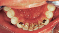
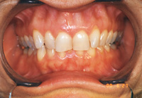
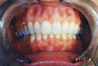
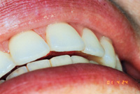
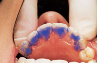
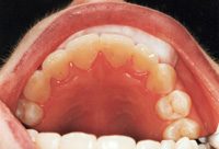
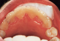
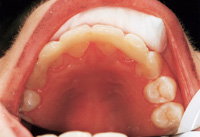
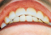
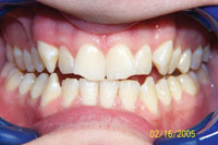
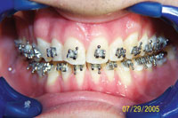
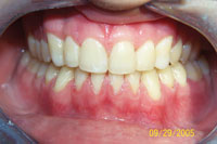
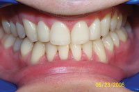
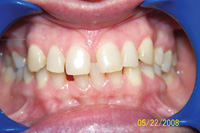
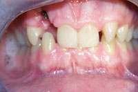
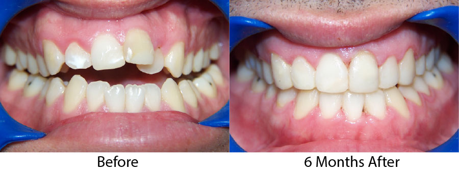
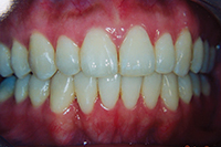
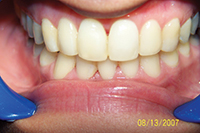
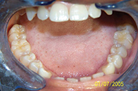
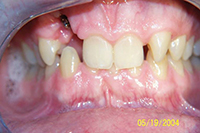
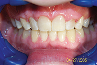
Case I

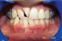
The patient presented with typical Class I crowding with aesthetics as the chief complaint. Rather than expand the arches into an unstable position prone to relapse in the adult patient, or reproximate lower incisors so much that they impinge on the gingival embrasures, it was decided to remove a lower incisor. The uppers were reproximated using a Brassler diamond disc and edges recontoured. The treatment was seven months and the patient was splinted afterward. Some molar supra-eruption occurred because of an anterior composite bite plane that relieved the deep bite and decreased the likelihood of further attrition in the anteriors.
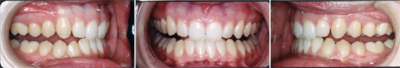
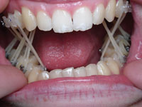
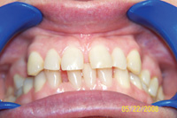
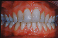
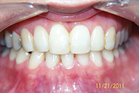


Case II

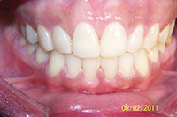
The patient was referred by a local dentist who had done simple orthodontics, but who was not willing to treat occlusal problems. The patient had crowding with a bilateral crossbite that was causing both anterior and posterior attrition at a young age, requiring orthodontics.

The crossbite was corrected through the use of cross-arch elastics from the lingual of the upper molars and bicuspids to the buccal of the lowers. Enamel reproximation made space to treat the anterior crowding. Upper and lower bonded Ribbond splints served to reinforce the bonded incisal areas caused by attrition. It also provided resistance to fracture, as the splints produce a greater bonded surface area and composite thickness. An upper posterior Hawley retainer prevented relapse of the posterior crossbite.
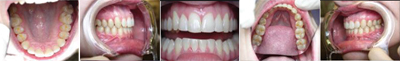
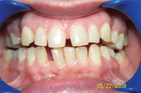
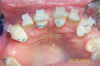
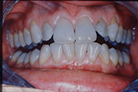
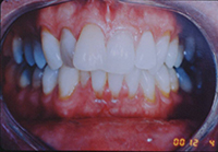
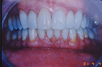


Case III

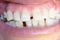
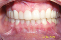
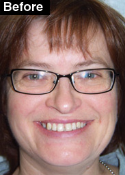
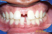
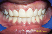
This patient presented with the chief complaint of a large diastema. She had advice from numerous orthodontists who expressed different opinions regarding how to correct this (because of her deep bite and lack of lower spacing), as well as reservations regarding the possibility of successful retention. At our consultation, it was explained to the patient that our plan would include:

  • Upper and lower anterior retraction and possibly lower enamel reproximation because of extra space on the uppers;
  • A fixed composite bite plane on #8 and #9 lingual to relieve the deep bite by causing posterior supra-eruption;
  • Possibly redistributing excess space to the distal of the upper canines to limit the retraction required; and
  • An upper splint, which would be required. Removable retention is unacceptable in these cases. Therefore slight overjet in the final result is planned to make space for the splint.


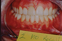
Per usual protocol, a prophy, bitewings, panoramic X-rays and restorative work were completed first. The patient’s treatment lasted five and a half months, with splinting and bleaching occurring on the final visit. At recall, the patient’s Ribbond splints were intact as she was not a bruxer. It is unlikely that this case would have succeeded without fixed retention.

It has been estimated that in 1970, only 5 percent of adults aged 18 or older sought consultations for comprehensive orthodontic treatment. In 1990, four times that number sought consultations for orthodontics.

Conclusion

Six-month adult cosmetic orthodontic treatment has a 60 percent acceptance rate among new patient consults in my practice, and post-treatment satisfaction is high. Many adults who undergo treatment have previously declined comprehensive treatment in other offices. Enamel reproximation, extraction of a lower incisor for space and limited occlusal change are among the modalities making this treatment unique and well accepted by patients. Offering clear or lingual appliances increases the patient’s cosmetic options. Treatment planning the orthodontic and restorative phases together facilitates patient understanding and communication, and delivers an outstanding cosmetic service. Patients with TMD, skeletal chief complaints, severe over/underjet, occlusal problems or very deviated midlines may opt for comprehensive treatment by an orthodontist. However, for the majority of adult patients with simply unaesthetic, crowded, spaced, functionally efficient and non-TMD dentitions, dentists should focus on the aesthetic chief complaint by performing conservative attenuated treatment in the general practice.

References

  1. Gottlieb EL. 1990 JCO study of orthodontic diagnosis and treatment procedures: results and trends. J Clin Orthod. 1991;24:145-56.
  2. Nattrass C, Sandy JR. Adult orthodontics—a review. Br J Orthod. 1995 Nov;22(4):331-7.
  3. Varela M, García-Camba JE. Impact of orthodontics on the psychologic profile of adult patients: a prospective study. Am J Orthod Denofacial Orthop. 1995 Aug;108(2):142-8.
  4. Lew KK. Attitudes and perceptions of adults towards orthodontic treatment in an Asian community. Community Dent Oral Epidemiol. 1993 Feb;21(1):31-5.
  5. Cochrane SM, Cunningham SJ, Hunt NP. Perceptions of facial appearance by orthodontists and the general public. J Clin Orthod. 1997 Mar;31(3):164-8.
  6. Proffit WR. Contemporary orthodontics. 2nd ed. St Louis: Mosby; 1993. p. 155.
  7. Little RM, Riedel RA, Engst ED. Serial extraction of first premolars—postretention evaluation of stability and relapse. Angle Orthod. 1990 Winter;60(4):255-62.
  8. McReynolds DC, Little RM. Mandibular second premolar extraction—postretention evaluation of stability and relapse. Angle Orthod. 1991 Summer;61(2):133-44.
  9. Weintraub JA, Vig PS, Brown C, Kowalski CJ. The prevalence of orthodontic extractions. Am J Orthod Dentofacial Orthop. 1989 Dec;96(6):462-6.
  10. O’Connor BM. Contemporary trends in orthodontic practice: a national survey. Am J Orthod Dentofacial Orthop. 1993 Feb;103(2):163-70.
  11. Paquette DE, Beattie JR, Johnston LE Jr. A long-term comparison of nonextraction and premolar extraction edgewise therapy in “borderline” Class II patients. Am J Orthod Dentofacial Orthop. 1992 Jul;102(1):1-14.
  12. Glenn G, Sinclair PM, Alexander RG. Nonextraction orthodontic therapy: posttreatment dental and skeletal stability. Am J Orthod Dentofacial Orthop. 1987 Oct;92(4):321-8.
  13. Begg PR. Stone Age man’s dentition. Am J Orthod. 1954;40:298-312.
  14. Sheridan JJ, Ledoux PM. Air-rotor stripping and proximal sealants. An SEM evaluation. J Clin Orthod. 1989 Dec;23(12):790-4.
  15. Sheridan JJ. The physiologic rationale for air-rotor stripping. J Clin Orthod. 1997;31:609-12.
  16. Peck H, Peck S. An index for assessing tooth shape deviations as applied to the mandibular incisors. Am J Orthod. 1972 Apr;61(4):384-401.
Read More

Anterior Retention With a Reinforced Composite Resin Splint After Cosmetic Orthodontic Treatment

Anterior Retention With a Reinforced Composite Resin Splint After Cosmetic Orthodontic Treatment

Originally posted on Dentistry Today.

It has become increasingly clear that making space in the crowded adult dentition by orthodontic expansion of the dental arch is prone to relapse.1,2 Moreover, the intercanine distance has actually been shown to decrease as early as mid-adulthood.1,3 Even cases treated to stability during late adolescence are prone to “late incisor crowding” by 34 years of age,4and the presence of third molars does not significantly contribute to this.5 Even though other skeletal factors and even facial bone dimensions may not significantly decrease until a later age, the influence of naturally decreasing intercanine distance on anterior dental aesthetics has been grossly underestimated. This basic misunderstanding of the early maturation of adult jaw dimensions has enormous repercussions for orthodontic treatment philosophy, as well as implications for the necessity of long-term retention.

Therefore, in cases where one must choose between slight contraction of the intercanine dimension through lower incisor extraction or expansion, the former may prove more stable. Anticipating “intercanine shrinkage” may help prevent future crowding years later when the doctor and patient have presumed stability, and retention has been discontinued. A cosmetic splint anchoring each anterior tooth individually serves to prevent this common and unaesthetic phenomenon of anterior orthodontic relapse.

Traditionally, orthodontists in the 1950s used a prefabricated metal bar fixed to only the canines for lower retention, with the rationale being the effects of arch expansion would be maintained until it was removed. Any incisor relapse would be inconsequential because it would just be an “aesthetic” consideration (Figure 1). Now that dentistry has accepted that the major reason adult patients seek treatment is aesthetics, we can properly address this essential aspect of retention. Some began affixing a customized bar with incisor pads or braided wire bonded to each incisor,6,7 which represented an improvement but still required placing metal in an aesthetic area. It was rationalized that the elastic property of a thin wire allowed physiologic mobility helpful in the periodontic patient.7 This is to be differentiated from the orthodontic patient with healthy periodontium, where the aim is to provide a window for bone and PDL fibers to reorganize with rigid reinforcement.

Also presented for periodontal patients have been reinforced composite splints using TMS pins8 and bondable reinforcement ribbon.9 With the boom in cosmetic dentistry and cosmetic orthodontics, the ribbon is proving useful for the stabilization of adult patients. Unlike the lingual bar this splint can be later removed incrementally as the patient desires.10 As 50% of relapse has been shown to occur in the first 2 years after orthodontic treatment,11 the splint should remain intact for longer than 2 years.

The main purpose of the splint is rigid fixation of the teeth. This immobilization, however, also accelerates the growth of supporting tissues, as the alveolus and PDL fibers can reorganize around the teeth in their new positions without interference from tooth mobility inherent in orthodontic treatment. In addition, this technique enables cosmetic augmentation of the final orthodontic result, as black triangular spaces, incisal discrepancies, or the lengthening of teeth can be achieved with more strength than free-standing incisal composites, which lack the thickness or support of a reinforcement material on the lingual aspect (Figures 2 and 3). Except for those few cases where the patient has a perfect orthodontic result and well-proportioned white teeth without any incisal defects, anterior bonding attached to and reinforced by the splint can greatly enhance the final aesthetic result.

If a maxillary splint is planned and the patient presents with overjet, the overjet should be preserved to allow space for the maxillary splint (Figure 4). This is in contrast to traditional orthodontic philosophy of complete elimination of overjet, even if the overjet represents the natural skeletal position. Skeletal changes cannot be permanently retained without surgery. Adult overjet, such as in a class 2, division 2 case (Figure 5), will be more stable if the overjet is maintained.

Figure 1. Lingual metal bar fixed only to the canines allows incisor relapse, which is not acceptable in cosmetic orthodontic patients. Figure 2. Class 2, division 2 before incisors are tipped forward giving overjet. Note attrition from deep bite on palatally tipped incisors.
Figure 3. After a 6-month treatment time with lingual braces, patient is splinted. Irregular incisors may be lengthened with more durability than with incisal composites not supported by a splint. Figure 4. Slight overjet in final result helps allow the necessary thickness for a durable maxillary splint.
Figure 5. Once completed, an adult class 2, division 2 case will result in overjet without surgery. Figure 6. Etching can include incisals should there be discrepancies that need correction.
Figure 7. Initial layer of composite should be a strong material and kept away from papillae. Figure 8. Splint-It! reinforcement material is placed into composite and cured.
Figure 9. Placement of addtional composite to cover reinforcement fibers. Figure 10. Occlusion is checked before final recontouring and polish.

SPLINTING STEPS

Step one. Complete enamel etching with recontouring on buccal and incisal for aesthetics, and on lingual if necessary to allow splint thickness with occlusion (Figure 6).

Step two. Bonding layer with composite is kept away from gingiva (Figure 7).

Step three. Two strips of Splint It! (Jereric/Pentron) or Ribbond (Ribbond Inc) reinforcement material are pressed into composite. Excess material is placed over reinforcement and cured (Figure 8).

Step four. Addition of final layer of composite (Figure 9).

Step five. Occlusion is checked preceeding recontouring embrasures with a Brasseler No. 8392-31 016F interproximal diamond and polishing bur (Figure 10).

SUMMARY

Even in the most stable types of orthodontic treatment, any relapse at all may be unacceptable cosmetically. Through the placement of a reinforced composite splint, the teeth can be held in position and more significantly recontoured, thus augmenting the final result. Subsquent splint removal can be done incrementally 3 to 5 years after placement as the patient desires.

Author’s Note: I was saddened to hear of the passing of Dr. John Witzig on December 3, 2001. Dr. Witzig was a true innovator who was not afraid to fight the tide of consensus in orthodontics. He brought many  people together in the field (I met my wife at his course). We all owe him a debt of gratitude, and he will be greatly missed. Thank you, John.


References

1. Bishara SE, Jakobsen JR, Treder J, et al. Arch width changes from 6 weeks to 45 years of age. Am J Orthod. 1997;111:401-409.

2. Rossouw PE, Preston CB, Lombar CJ, et al. A longitudinal evaluation of the anterior border of the dentition. Am J Orthod Dentofaciai Orthop. 1993;104:146-152.

3. Sinclair PM, Little RM. Maturation of untreated normal occlusions. Am J Orthod. 1983;83:114-123.

4. Bondevik O. Changes in occlusion between 23 and 34 years. Angle Orthod. 1998;68:75-80.

5. Harradine NW, Pearson MH, Toth B. The effect of extraction of third molars on late lower incisor crowding: a randomized controlled trial. Br J Orthod. 1998;25:117-122.

6. Becker A, Goultschin J. The multistrand retainer and splint. Am J Orthod. 1984;85:470-474.

7. Oikarinen K. Comparison of the flexibility of various splinting methods for tooth fixation. Int J Oral Maxillofac Surg. 1988;17:125-127.

8. Rosenberg ES, Garber DA. A temporary-permanent splint. Refuat Hapeh Vehashinayim. 1979;28:27-30,33-37.

9. Ferreira ZA, de Carvalho EK, Mitsudo RS, et al. Bondable reinforcement ribbon: clinical applications. Quintessence Int. 2000;31:547-552.

10. Sheridan JJ. Incremental removal of bonded lingual retainers. J Clin Orthod. 1988;22:116-117.

11.Kuijpers-Jatman AM, Al Yami EA, van’t Hof MA. Long-term stability of orthodontic treatment. Ned Tijdschr Tandheelkd. [in Dutch] 2000;107:178-181.

Read More

Occlusal Change Through Orthodontics in TMD Patients

Occlusal Change Through Orthodontics in TMD Patients

Originally posted on Dentistry Today.

Although some claim that occlusion has little effect on a healthy TMJ and is not generally a causal factor in TMD,1,2 many have made emphatic claims to the contrary.3 Many orthodontic practices are positioned in the marketplace as providing proven treatment for TMD, yet some patients seem to experience TMD as a consequence of orthodontic treatment.
While existing literature reports that orthodontics can both helpand worsen TMD, this paper describes 2 cases where TMD relief was successfully achieved through orthodontic therapy. More specifically, these cases lend credence to the theory that increasing the vertical dimension5 and removing retrusive forces on the mandible may help recapture the disc that can be displaced by over-closure of the mandible.

CASE 1

Figure 1. Photos of patient when she presented for fixed orthodontic treatment. She had been wearing a removable splint and was asymptomatic but splint dependent.

Case 1 describes treatment that utilized a hard acrylic, flat-planed splint to alleviate TMD symptoms of pain, popping, and clicking by advancing the mandible and increasing vertical dimension. The patient was “splint dependent” but symptom-free at the stage she was transferred for orthodontic treatment (Figure 1). The pain returned whenever she was not wearing her splint for consecutive days because she returned to an “over-closed” position. Once orthodontic treatment commenced, the splint was reduced incrementally, allowing teeth to supra-erupt. This was done sequentially until the natural occlusion mimicked the patientís occlusion with the splint. It was reduced from the posterior forward, allowing the second molars to supra-erupt in a controlled fashion. It was also sequentially reduced in thickness. Mobility from the orthodontics facilitated this occlusal setting. Three distinct aspects of the patientís occlusion were changed, which helped provide TMD relief:

(1) The maxillary incisors were flared labially with treatment. Lingually inclined  lower incisors translate occlusal force into a retrusive direction as the patient closes, especially during protrusion. This was eliminated as labially inclined upper and lower incisors deliver chewing force in a more vertical direction into the alveolar bone, decreasing the tendency of the mandible to be pushed backward and minimizing disc trauma.

(2) Similarly, the incisors had greater vertical overlap initially. This compounded the problem caused by the retroclined position, as the entire facial surface of the lower incisors was acting as a receiving surface for ìpoundingî by the maxillary incisors. The posterior dentition better tolerates this vertical chewing force.

Figure 2. Cross-arch vertical elastics used to bring posterior extrusion without tipping. The splint was reduced incrementally.

(3) The molar extrusion and improved interdigitation, in conjunction with occlusal adjustment, provided a more stable posterior occlusion. This offers better protection against retrusive slides in centric and during mastication, which can further exacerbate TMD. Molar extrusion achieved using cross-arch elastics (Figure 2) from the buccal of the upper teeth to the lingual of the lowers as well as lingual of the upper teeth to the buccal of the lowers served to extrude the posteriors with greater control and no buccal-lingual tipping.

Figure 3. Occlusion after removal of braces.

Although the causal factors of TMD are often a mystery, this case demonstrates that eliminating obvious and severe occlusal abnormalities through splint therapy and gradually through or-thodontics may provide TMD relief and minimize occlusal wear as the traumatic occlusion is eliminated (Figure 3). Two years after treatment, the patient was orthodontically stable and symptom-free.

CASE 2

Figure 4. Patient’s occlusion before treatment. Figure 5. Progress at 5 months.
Figure 6. “After” photo with upper and lower teeth splinted and incisals restored. Figure 7. Eleven-month recall.

The second case shows a patient who had bilateral TMJ clicking and tinnitus. He had second molar occlusion only, a constricted maxillary arch, occlusal trauma, and wear (Figure 4).
The patient wore posterior cross-arch elastics from the lingual of the maxillary posteriors to the buccal of the mandibular posteriors to achieve proper intercuspation and bilateral, evenly distributed tooth contacts, as a posterior cross-bite has been associated with TMD.6 The upper posteriors were stabilized with a Hawley retainer. The upper and lower anteriors were stabilized with lingual Ribbond splints (Ribbond) canine to canine.
This effectively stabilized rotated teeth (in conjunction with a fiberotomy) and provided proper resistance form to the restored incisal composites, necessary because of the previous occlusal trauma (Figures 5 and 6). The incisal edges became much more durable once connected to the splint because of increased thickness. The TMJ, occlusion, and restored incisal surfaces were all stable at recall (Figure 7).

CONCLUSION

While TMD is often a mystery and is even seen in many normal occlusions, frequently other factors7 exist, such as a history of trauma, bruxism, or degenerative joint disease of a systemic nature. However, these 2 cases show at least one obvious and proximate cause for their TMD, which is an unstable occlusion.
Acute inflammation can be mitigated through ice, NSAIDS, and splint therapy until subsequent inevitable exacerbations occur. Definitive treatment through permanent occlusal change sometimes is the only hope for these patients, and is still not a panacea if disc damage has occurred or if occlusal abnormalities are not corrected.
While all aspects of orthodontic TMD treatment have not been substantiated in the literature, providing the patient with a stable, evenly distributed occlusion with correct buccal-lingual molar and nonretrusive incisor relationship, as well as providing an increased vertical dimension, may be a good place to focus in treating this elusive problem.

References

  1. Gesch D, Bernhardt O, Kirbschus A. Association of malocclusion and functional occlusion with temporomandibular disorders (TMD) in adults: a systematic review of population-based studies. Quintessence Int. 2004;35:211-221.
  2. Gesch D, Bernhardt O, Mack F, et al. Association of malocclusion and functional occlusion with subjective symptoms of TMD in adults: results of the Study of Health in Pomerania (SHIP). Angle Orthod. 2005;75:183-190.
  3. Reinhardt R, Tremel T, Wehrbein H, et al. The unilateral chewing phenomenon, occlusion, and TMD. Cranio. 2006;24:166-170.
  4. Henrikson T, Nilner M, Kurol J. Signs of temporomandibular disorders in girls receiving orthodontic treatment. A prospective and longitudinal comparison with untreated Class II malocclusions and normal occlusion subjects. Eur J Orthod. 2000;22:271-281.
  5. Hisano M, Ohtsubo K, Chung CJ, et al. Vertical control by combining a monoblock appliance in adult class III overclosure treatment. Angle Orthod. 2006;76:226-235.
  6. Thilander B, Rubio G, Pena L, et al. Prevalence of temporomandibular dysfunction and its association with malocclusion in children and adolescents: an epidemiologic study related to specified stages of dental development. Angle Orthod. 2002;72:146-154.
  7. Clark GT. Etiologic theory and the prevention of temporomandibular disorders. Adv Dent Res. 1991;5:60-66.
Read More

A Six-Month Orthodontic Solution to Space Closure and Bite Collapse

A Six-Month Orthodontic Solution to Space Closure and Bite Collapse

Originally posted on Dentistry Today.

For patients who exhibit missing posterior teeth, bruxism, and a concomitant loss of vertical dimension often commonly occurring with anterior flaring and spacing1 (Figures 1 to 3); mainstream treatment consists of 1.5 to 2 years of orthodontic treatment to retract the anteriors and re-establish the collapsed vertical dimension. This is usually followed by removable retainer wear. It is important to restore the missing posterior support,2 and the patient should be given implants or bridges afterward.

CASE REPORT

Figures 1 and 2. Note palatal occlusion.
Figure 3. Note attrition.
Figures 4 and 5. Composite bite plane on teeth Nos. 6, 8, 9, and 11 intruded the anteriors and allowed passive eruption of posteriors.
Figure 6. After bridge cementation. Additional whitening procedures were recommended.

A patient who came to our general practice was given this treatment plan by 2 previous dentists with specialists in their offices. Eager to seek other alternatives, she presented for attenuated orthodontic and restorative treatment.
Treatment consisted of short-term, 6-month, fixed-orthodontic treatment by retracting the incisors to their original position before they migrated forward. The collapsed vertical dimension was increased through use of an anterior fixed composite bite plane. This is a flat-planed composite bite plane bonded to the lingual of the upper central incisors3 (Figures 4 to 6), prohibiting full closure. Through lack of posterior occlusion, within 3 to 4 months the posterior teeth exhibited significant passive supra-eruption, even without posterior vertical elastic wear (which may be used as an option to accelerate the process). At the same time, the incisor region is intruded through chewing. This occurs throughout the entire anterior region, as the teeth are essentially “splinted” through the orthodontic wire. In this way, even teeth without the composite bite plane are intruded. The ratio of posterior extrusion to anterior intrusion has been shown to be approximately 60:40.4

DISCUSSION

Figure 7a and 7b. Before and After.

Secure retention is an essential aspect of this case. Removable retainers are inadequate, as even slight space relapse will be cosmetically obvious; this is likely in an adult patient with fully formed dental arches and some bone loss.5,6 In addition, our practice occupies a niche in treating adults through short-term cosmetic orthodontics,7 and this demographic desires retention that is aesthetic. Furthermore, treatment is orthodontic in these cases and not orthopedic, so the results are less stable, thus requiring fixed retention. A lingual composite splint (Ribbond [ribbond.com]), where composite covers most of the tooth’s lingual aspect and can overlap onto the buccal aspect, is preferred. This can serve to augment small teeth, change shape and width by enhancing line angles, fill chips, and restore surfaces with attrition.8
In conjunction with the orthodontic space closure, posterior support must be provided, as the splint will fracture without posterior protection and incisor flaring will return.9 The increased vertical dimension would also be lost, since the posteriors would intrude. If implants are part of this plan, they should be placed before or during orthodontic treatment, not after. This case utilized 3 fixed bridges, helping to correct some mesial drift which may be caused by transseptal fiber contraction.10Temporary bridges were inserted the day the braces were removed, and the splints were placed. Permanent impressions were taken one month later to allow for gingival healing and minor occlusal settling (Figures 7a and 7b).

CONCLUSION
This treatment approach shows a rapid, straightforward solution for this common functional and aesthetic dental problem, which is frequently treated with a more complicated long-term plan, often prone to relapse.

CONTACT RAPID BRACES


References

  1. Kelly JT Jr. A multidisciplinary approach to restoring posterior bite collapse. Compend Contin Educ Dent. 1997;18:483-485,488-490.
  2. Reshad M, Jivraj S. The influence of posterior occlusion when restoring anterior teeth. J Calif Dent Assoc. 2008;36:567-574.
  3. Georgaklis CC. Alternative orthodontic treatment for adult crossbites and overbites. Dent Today. 2001;20:60-63.
  4. Lei Y, Zhang S. Clinical study on the orthodontic treatment of deep overbite with bite plane [in Chinese]. Hunan Yi Ke Da Xue Xue Bao. 1998;23:465-466.
  5. Brunsvold MA. Pathologic tooth migration. J Periodontol. 2005;76:859-866.
  6. Martinez-Canut P, Carrasquer A, Magán R, et al. A study on factors associated with pathologic tooth migration. J Clin Periodontol. 1997;24:492-497.
  7. Georgaklis CC. Six-month adult aesthetic orthodontic treatment. Dent Today. 1999;18:110-113.
  8. Georgaklis CC. Anterior retention with a reinforced composite resin splint after cosmetic orthodontic treatment. Dent Today. 2002;21:54-57.
  9. Greenstein G, Cavallaro J, Scharf D, et al. Differential diagnosis and management of flared maxillary anterior teeth. J Am Dent Assoc. 2008;139:715-723.
  10. van Beek H. Dissertation 25 years later. 1. Mesial drift of teeth by occlusal forces [article in Dutch]. Ned Tijdschr Tandheelkd. 2004;111:48-51.
Read More

Adult Orthodontics and a Post-Treatment Bonded Splint: A New Cosmetic Subspecialty

Adult Orthodontics and a Post-Treatment Bonded Splint: A New Cosmetic Subspecialty

Originally posted on Dentistry.com

As the field of cosmetic dentistry is still rapidly evolving, we have not yet integrated the various specialties to provide seamless care in a case that requires multiple disciplines. For example, a patient needing orthodontics, crown and bridge or implants, gingival recontouring, orthodontic retention, and bonding will normally be cross-referred between their GP, periodontist, orthodontist, and perhaps an oral surgeon1,2 (4 doctors). While specialists normally provide the highest level of care, there exist certain cases where a “cosmetic subspecialist” may be best suited to create a final result that is harmonized in concept, proportion, and materials.
The following case would often be treated with orthodontics and retainers, and then relapse because of the unusual nature of the case and high tendency to relapse with removable retainers3 regardless of the duration of the orthodontics. The relapse in the anterior segment in adult patients is especially high.4 This patient had 3 missing anterior teeth as well as an impacted canine (Figure 1). Most orthodontists are not accustomed or trained to incorporate significant bonding and reshaping into their treatment plans, so the missing lateral incisors spaces would usually be opened up for implants or 6 units of crown and bridge to provide traditional canine guidance. Yet, there is insufficient bone for an implant for No. 7, and there are alternative treatment options that are simpler, far shorter in duration, and less expensive. Nontraditional thinking is required in this case, especially from an orthodontic perspective, as ideal treatment is most likely impossible.

CASE REPORT (FIGURE 1)

Figure 1. Patient is missing 3 lateral incisors. Tooth No. 6 was fully impacted in bone before surgical exposure, and was used to substitute for the missing tooth No. 7 through extrusion, mesialization, reshaping, and bonding. Figure 2. Bonding and reshaping was also done to the enamel and gingiva of the bicuspids to make them into canines. The front 6 teeth were splinted and bonded. This case has no implants or bridges.

The patient had been to 3 orthodontists and was looking for treatment alternatives, as all 3 offered 2- to 3-year treatment plans with no promise of a satisfactory result because of bone issues surrounding tooth No. 6, among other things. The patient was not willing to crown all his anterior teeth, which had also been proposed. Our treatment plan involved surgical exposure and super-eruption of tooth No. 6, then making teeth Nos. 6 and 11 into lateral incisors through mesial movement and bonding, making teeth Nos. 5 and 12 into canines, minimizing the lower canine cusps, and connecting all anteriors with a Ribbond/composite splint for stability as well as to support the extra bonding that would be necessary (Figure 2).5
Instead of dividing the treatment by “specialty” and fragmenting the plan with 3 providers, in this way the case may be managed by one practitioner who is experienced in performing splinted retention with concurrent bonding. I previously wrote of a porcelain pontic veneer placed over a Ribbond splint used for orthodontics.6 This case shows how the splint can support significant bonding and provide needed fracture resistance to canines, which are made into laterals, and bicuspids which are made into canines. Equally important is the required fixation, as any relapse would reveal their deficient anatomy and ruin the “camouflage” effect of the bonding. Most orthodontic cases have some degree of relapse, and that would be unacceptable in cases such as this.
This treatment reflects a paradigm shift. Instead of providing a traditional orthodontic result by adding prosthetic lateral incisors, other teeth are moved shorter distances and bonded, providing a faster, simpler, and less expensive result. This is preferable for many patients who are more interested in avoiding gaping holes in their smile than committing to a 2-year treatment plan with endosseous implants. While patients are made aware of treatment alternatives, specifically what will not be provided in this plan, they overwhelmingly choose a method that resolves their chief aesthetic complaint. They are generally satisfied years at recall as well. Should they choose to crown or veneer this result in the future when more stability has been achieved, that option is still available to them.

CONCLUSION


Reshaping teeth via splinting and bonding is a practical, aesthetic alternative which addresses the high incidence of relapse still seen in orthodontics today. Simultaneously, this can serve to resolve some difficult and unusual aesthetic predicaments.


References

  1. Chadroff B. The interdisciplinary approach to implant dentistry. Gen Dent. 2004;52:321-326.
  2. Spear FM, Kokich VG, Mathews DP. Interdisciplinary management of anterior dental esthetics. J Am Dent Assoc. 2006;137:160-169.
  3. Hirschfelder U, Hertrich K. The treatment of deep bite in adults. Fortschr Kieferorthop. 1990;51:36-43.
  4. Lang G, Alfter G, Goz G, et al. Retention and stability—taking various treatment parameters into account. J Orofac Orthop. 2002;63:26-41.
  5. Kokich VO Jr, Kinzer GA. Managing congenitally missing lateral incisors. Part 1: Canine substitution. J Esthet Restor Dent. 2005;17:5-10.
  6. Georgaklis CC. Anterior retention with a reinforced composite resin splint after cosmetic orthodontic treatment. Dent Today. Jan 2002;21:54-57.
Read More

6 Month Braces: How Is It Possible?

6 Month Braces: How Is It Possible?

Get straight teeth in just six months with Dr. Georgaklis’ Rapid Braces treatment. He specializes in clear braces or invisible braces for adults and promises to give you your best smile.

rapid braces Boston

Adults who need braces shouldn’t have to suffer for a long time period with metal brackets on their teeth — let Rapid Braces be the solution for you! This Brookline dental office is the only office to complete 90% of cases in six months or less. Adults can wear clear braces or invisible braces and see results faster! These also are a better alternative to Invisalign, as they control your teeth with more force and take less the time.

How does Dr. Georgaklis do it?

First, he makes sure that every patient at his Boston area dental office gets personalized treatment the moment they walk through the door. He schedules each patient for longer visits, allowing him to get more done during one appointment. Most orthodontists see their patients for five to fifteen minutes, while Dr. G sees his patients for an hour.

Dr. Georgaklis uses his professional experience to mix cosmetic dental practices with orthodontic techniques to give his patients straight teeth fast.

In order to treat patients within six months, this Boston area dentist uses special techniques like:

  • using special wires that don’t fatigue
  • sanding in between the teeth
  • not removing any teeth
  • reshaping gums and teeth
  • not changing the bite or profile unless needed (although this can often times be done in six months)
  • straightening front teethadult braces

These procedures give you a perfect smile with proportionate and straight teeth! After six month braces, Dr. Georgaklis fits his patients with a fixed retainer behind the teeth. This lingual retainer prevents movement, giving you straight teeth for life!

To learn more about the Six Month Braces treatment, click here. You can book your free consultation today!

Read More

Difficult Case Types: Part 2, A Discussion of Adult Short-Term Orthodontics

Difficult Case Types: Part 2, A Discussion of Adult Short-Term Orthodontics

Clear or Invisible braces for adults.

Originally posted on Dentistry Today.

INTRODUCTION 
In part 1 of this article, I discussed the evolution and rationale of short-term orthodontics (STO); and how it dovetails with aesthetic dentistry and other disciplines to provide treatment by one provider in a coordinated and timely manner. I briefly discussed issues involving treating patients who present with bruxism, patients who have unrealistic cosmetic demands or obsessive-compulsive disorder, and those personality types who wish to alter or control their treatment plan. Though STO is oriented toward the patient’s aesthetic chief complaint, we must still limit the patient’s involvement in the treatment planning and smile design to meet “real world” expectations.
At the initial consult, it is helpful to provide cosmetic orthodontic patients with before and after pictures of what they may expect, along with a list of what can and cannot be achieved. All common significant questions should be addressed on one’s Web site, in addition to a consultation photo book given to new patients before seating. Actual pictures of one’s own work can be a fair representation of what to expect, providing an honest and direct dialogue that can be very beneficial to both the patient and the provider.
The case types discussed in the second part of this article include interesting aspects of transfer cases, the judicious use of enamel reproximation, cases with particular retention needs, cases with temporomandibular disorders (TMD), large tongues, profile problems, and a complex multidisciplinary case. These difficult case types offer an opportunity to present pearls that can enhance treatment for both STO and conventional orthodontics alike. The greater focus and time per visit required for STO (I prefer one-hour visits) also bring greater reward and satisfaction for both the patient and the provider.

CASE TYPE IV: TRANSFER CASE WITH TIPPED POSTERIOR TEETH
While some believe that they can nonsurgically expand the dental arch through orthodontics in an adult patient, it has been proven that “expansion” (crown tipping in an adult) at past the age of 13 years is not significant, and it is prone to relapse.1Posterior crowns tip to the buccal without significant root translation resulting in an unaesthetic and unstable result also prone to gingival recession.

Figure 1. Patient first came into our office with teeth tipped outward through use of removable expansion appliances. There was sufficient space for alignment, but the roots were in unstable positions with crowns tipped to the buckle. Figure 2. A stable result must maintain arch circumference in an adult patient with the teeth in cortical bone to prevent inward collaspe post-treatment.
Figure 3. Patient presented with narrow incisors due to previous treatment that relied too heavily upon enamel reproximation and scarificed tooth proportion and aesthetic outcome.

Patient in Figure 1 was referred by an orthodontist in Los Angeles for lingual orthodontics. He expanded her for one year with Crozat appliances, leaving sufficient space to align the teeth (Figure 1). At this point, she moved to Boston, where we commenced lingual orthodontic treatment which proceeded smoothly. Brackets were removed with an aesthetic result. However, in the months after completion, the arch form and tooth roots continued to collapse inward. Expansion had spread the teeth laterally into an unstable position outside the cortical bone. The patient needed a brief course of retreatment with enamel reproximation which yielded a far more stable result which has been maintained well (Figure 2).

CASE TYPE V: OVERUSE OF ENAMEL REPROXIMATION
While the previous case showed an under-reliance on enamel reproximation, this case shows overreliance on it. Lack of flexibility and overreliance on any one treatment modality has its perils, though. The patient in Figure 3 was looking for retreatment despite the fact that her teeth were straight. In order to achieve an ideal occlusion nonextraction by the treating orthodontist, the teeth had been interproximally reduced to the point that they were unaesthetic, lacked embrasure space, and were not self-cleansing. This resulted in unaesthetic tooth proportions and perpetually inflamed papillae. Minor alignment was done along with recontouring. The teeth were shortened to establish better proportion, and embrasure spaces were opened to allow better self-cleansing.

CASE TYPE VI: SPECIAL RETENTION NEEDS; ADULT CLASS II, DIVISION 2; LARGE DIASTEMA, SEVERE ROTATION 
The Class II, Division 2 is a common type of crowding where the upper centrals tip palatally and the laterals flare labially (Figure 4). Aesthetically conspicuous, it is usually a simple case to align dentally with enamel reproximation. These patients do not usually have a profile problem needing orthognathic surgery. As they are fully grown adult patients, skeletal change and complete overjet correction is not usually possible nonsurgically, so the upper central incisors will always tend to relapse palatally. Therefore, this is an ideal case for maxillary lingual splinting of teeth Nos. 7 to 10 or teeth Nos. 6 to 11. Slight overjet allows a durable splint to be placed out of occlusion in a case type that would otherwise be very prone to relapse. Recognizing the instability of cases that have a skeletal component is essential, and this patient’s aesthetics are basically identical today to the result (Figure 5), 12 years after completion, with no noticeable relapse due to her upper and lower lingual fiber-reinforced composite splints (Ribbond).

Figure 4. Adult Class II, Division 2 is very prone to relapse. Figure 5. Splinted result maintained well (at 12-year recall).
Figure 6. Large diastema needing fixed retention. Figure 7. After short-term orthodontics (STO) with splinting.
Figure 8. The 3.5-year recall with fiber-reinforced composite (FRC) (Ribbond) splints. Figure 9. Severely rotated incisor.
Figure 10. This rotation could never be maintained without a splint. Figure 11. Four-year recall with maxillary FRC splint.

Large diastema cases (Figures 6 to 8) also have special retention needs (a maxillary splint), as do severely twisted teeth (Figures 9 and 10). Though it requires overjet be left in the final result, the maxillary splint provides excellent retention, though it can require maintenance. Removable retainers would almost surely fail to retain these particular tooth movements. However, with the maxillary splint, the excellent results were well-retained in both cases at the 3- and 4-year recalls (Figures 8 and 11). Few orthodontists finish cases with the overjet needed to allow for placement of a maxillary splint.

CASE TYPE VII: Temporomandibular Disorder 
This patient was a bruxer whose crowding and anterior recession were worsened by bruxing forward, causing anterior displacement of an upper central incisor (Figure 12). As a prominent cosmetic dentist, he came to Boston for rapid cosmetic orthodontics. The alignment proceeded smoothly with one exception: I allowed the likable dentist-colleague to limit my enamel reproximation in the lower arch. Therefore, my ability to retract the lower incisors and establish sufficient overjet also became limited. Parafunction usually ceases at the beginning of orthodontic treatment, but then returns once the teeth are no longer sore. Once the parafunctional bruxing returned, the upper central (that now had been retracted back) caused a more retrusive and limiting anterior guidance on the mandible (Figure 13). The new incisal guidance brought less freedom of the mandible during bruxing, pushing it backward, so disc compression and tinnitus followed.

Figure 12. Bruxer, before STO, with protruded tooth No. 8 from bruxism. Figure 13. Bruxer, after STO, with normal incisor occlusion.

Our typical treatment method of leaving overjet avoids any retrusive incisor contact on the mandible, and avoids TMD sequelae. The lack of tight anterior coupling in my finished orthodontic cases accounts for the fact that I rarely see TMD in my patients after STO—a remarkable statistic, especially considering occlusal change is not the primary treatment focus. One must be very cautious when leaving a case with the incisors tightly coupled together in occlusion, as any lower incisor relapse or change in jaw position forward may cause disc compression and the pain that may or may not have been poresent beforehand.

CASE TYPE VIII: LARGE TONGUE 
Patients with a large tongue often have anterior spacing. The patient’s tongue in Figure 14 already fills the space available and goes to the lingual surfaces of the teeth. While the anterior spacing can be reallocated distal to the canines, the incisors cannot be retracted and maintained inside the neutral zone with long-term stability. The tongue pressure will push the teeth forward unless tongue reduction has occurred. In such cases, we always explain to our patients at the initial consult that space will be redistributed distally to maintain an incisor position that is in harmony with the tongue, instead of a retracted incisor position when the tongue will not allow them to be maintained and would cause relaspe.

CASE TYPE IX: PROTRUSION WITH UNAESTHETIC PROFILE
There is no STO solution for cases with an unaesthetic facial profile and lip incompetence. This case needed bicuspid extraction because the amount of upper incisor retraction required cannot be done with enamel reproximation alone (Figures 15 and 16).

Figure 14. Large tongue prohibited the retraction of incisors. Figure 15. Bicuspid extraction case with lip incompetence.
Figure 16. After bicuspid extraction treatment. Figure 17. Surgical case that requires a referral to the surgeon-specialist team.
Figure 18. Canine substitution needed for missing upper lateral incisors. Figure 19. Final result; bonded and splinted upper canines and bicuspids without any bridgework or implants.

Even more involved, the skeletal case in Figure 17 clearly needs orthognathic surgery.

CASE TYPE X: MULTIDISCIPLINARY CASE
This case cannot be done with orthodontics alone (Figures 18 and 19). Treatment involved surgical exposure and bringing down impacted canines throughout one year (still considered STO due to the complexity of the case), as well as splinting with reshaping and bonding. Canine substitution was done for the missing upper laterals incisors. With some creative thinking, this patient avoided any bridgework or implants as this result was achieved solely with orthodontics and bonding. Most patients enjoy a result with greater simplicity, stability, and predictability, while eliminating implant surgery and minimizing treatment time and expense. This type of thinking can bring people back to dentistry, especially adult patients like this with aesthetic problems who have not sought out care sooner due to obstacles inherent in a conventional and more involved treatment plan. Patients opt out of 2-year treatment when there is a shorter plan with proven results.

CONCLUSION
As dentists, we have a myriad of responsibilities that can make dentistry complex as well as rewarding. Diagnosis and treatment planning, patient management, and retention protocol all vary with a need to understand and accommodate each patient’s teeth and character. Comprehensive 2-year orthodontics may better address more complex cases, but there is also a demand for more rapid orthodontic treatment for the typical adult cosmetic cases.
We must always remember that elective cosmetic dentistry of any type often comes with a human dimension of personal preferences that is often distinctive. These preferences must be understood, addressed, and ideally, satisfied, within the parameters of a healthy and stable long-term result.


Reference

  1. Bishara SE, Jakobsen JR, Treder J, et al. Arch width changes from 6 weeks to 45 years of age. Am J Orthod Dentofacial Orthop. 1997;111:401-409.
Read More

Expert Boston Dental Care

Rapid Braces in Brookline provides expert Boston dental care to patients throughout the area. The main goal at Rapid Braces is to give patients perfect smiles that they can feel confident to show off to the world. Dr. Georgaklis likes to use a gentle dental treatment approach that is designed to meet each patients unique needs. The staff at Rapid Braces knows that your time is valuable and works to be as efficient as possible to minimize the amount of time that you need to be in the office. A healthy mouth starts with high-quality dental treatment.

We offer a number of different Boston general dentistry services at the Rapid Braces Brookline office. Our expert Boston dental treatment usually starts with a cleaning. At this initial meeting we can see the exact condition your mouth is in and put together a dental plan that will be best for you moving forward. Dental treatment in Boston can get expensive and we strive to offer treatment plans that will keep you healthy and keep money in your pocket. Rapid Braces will never recommend a complex dental procedure when a simpler one to get the same results.

expert boston dental care

The entire staff at Rapid Braces makes it their goal to give patients the healthiest possible mouth. Keeping your mouth and teeth clean is incredibly important to your overall health. Our office offers a number of cutting edge Boston dental services so we can meet all of our patients’ needs. After your initial dental examination we will address any urgent issues first and then work to create a plan that will give you a healthy mouth for life. Rapid Braces aims to create Boston dental treatment plans that are suitable for each individual and get fantastic results.

A comprehensive dental cleaning is only the start of treatment at Rapid Braces. There are a number of other dental services available to give patients perfect smile as well. We offer state of the art care in esthetic composite fillings, root canals, lifelike dental crowns and deep scalings. Dr. G. has run Boston’s best dental practice for almost 25 years and has given countless patients perfect teeth and healthy mouths. Call today to schedule your initial dental examination and get started with Boston’s best dental care.

Read More